Figure 3.
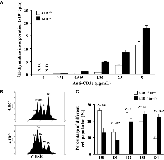
Hyperproliferation of 4.1R−/− T cells. (A) [3H] thymidine incorporation of CD4+ T cells. CD4+ T cells (105 per well) purified from lymph nodes of 4.1R+/+ mice or 4.1R−/− mice were stimulated with various concentrations of plate-bound anti-CD3ϵ as indicated. Proliferation was determined by [3H] thymidine uptake. The experiments were preformed for 6 times, and the data shown represent the mean value of triplicate samples from 1 experiment. (B,C) In vitro T-cell expansion. T-cell expansion in vitro was assessed by CFSE dilution as described in “In vitro T-cell expansion using CFSE.” Note fewer cells in D0 (undivided) and D1 (1 division) and more cells in D3 (3 divisions) and D4 (4 divisions) of 4.1R−/− CD4+ T cells compared with 4.1R+/+ CD4+ T cells, showing faster division of 4.1R−/− CD4+ T cells.

