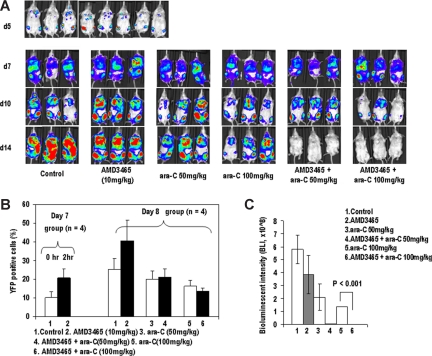
Figure 3.
AMD3465 induces mobilization of A20 cells in vivo and enhances antitumor effects of ara-C. (A) A20-luc/YFP cells were injected IV into BALB/c mice, and bone marrow engraftment was confirmed by bioluminescence imaging on day 5 (top row). Mice were injected with ara-C, AMD3465, or ara-C plus AMD3465 on day 7 and 8 at the indicated doses described in “A20-luc/YFP leukemia murine model.” On days 5, 7, 10, and 14, mice were imaged after D-luciferin injection. Serial images of 3 representative mice are shown on day 7, 10, and 14. (B) AMD3465 was administered on day 7 after tumor cell injection, and percentages of circulating A20-luc/YFP cells in peripheral blood before and after 1 hour of AMD3465 were examined by flow cytometry (left panel). Percentage of circulating A20-luc/YFP-positive cells in control mice, in mice mobilized with AMD3465, or in mice treated with ara-C ± AMD3465 was detected by flow cytometry on day 8 after tumor cell injection (right panel). (C) Bioluminescence imaging results on day 14 were averaged from the peak light-emitting exposure from each group and displayed as photons per second. Error bars represent the SEM of each group.

