
Figure 1. Renin Immunoreactivity in Cortical and Medullary Collecting Ducts of Rat Kidneys.
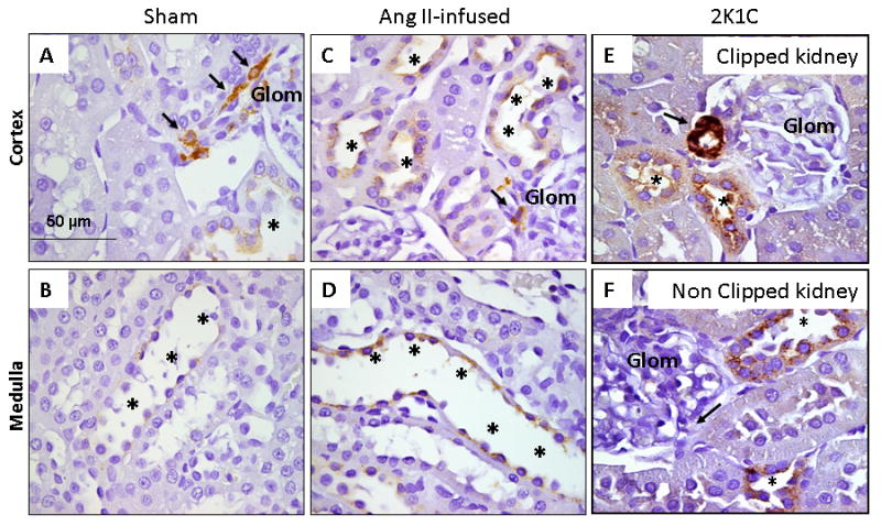
Panels A-F show the cortex (Panels A, C, and E) and medulla (Panels B and D) of kidney sections (3 μm) with specific renin immunostaining in sham rats (panels A-B), chronic Ang II-infused rats (panels C-D) and 2K1C Goldblatt hypertensive rats (panels E-F). Arrows show renin immunoreactivity in juxtaglomerular cells localization (DAB chromogen) in a sham (panel A), chronic Ang II-infused rat (panel C) and in the clipped kidney (panel E) and non-clipped kidney (panel F) from Goldblatt rats. Higher renin immunoreactivity (asterisks; DAB chromogen) are shown in the collecting ducts of the renal cortexes of chronic Ang II-infused rats (panel C) and both, clipped (panel E) and non-clipped (panel F) kidneys relative to sham kidney section (panel A). Glom: Glomerulus.