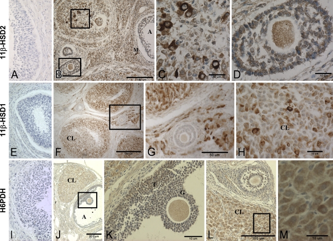
Figure 4.
Photomicrographs of immunohistochemical detection of 11β-HSD1, 11β-HSD2, and H6PDH in a rat ovary. (A,E,I) Negative controls for 11β-HSD1, 11β-HSD2, and H6PDH, respectively. (A–D) 11β-HSD2. (C,D) Higher magnifications of areas boxed in B. The follicle in upper left of B appears to be atretic and includes several active macrophages. (E–H) 11β-HSD1. (G) Higher magnification of boxed area in F. (H) Higher magnification of a corpora lutea showing intense 11β-HSD1 immunoreactivity in a few cells. (I–M) H6PDH. (K,M) Higher magnifications of boxed areas in J and L, respectively, showing the oocyte with preovulatory cumulus cells (K) and corpora lutea (M). A, follicular antrum; CL, corpora lutea; T, theca cells; G, granulosa cells; M, macrophage.

