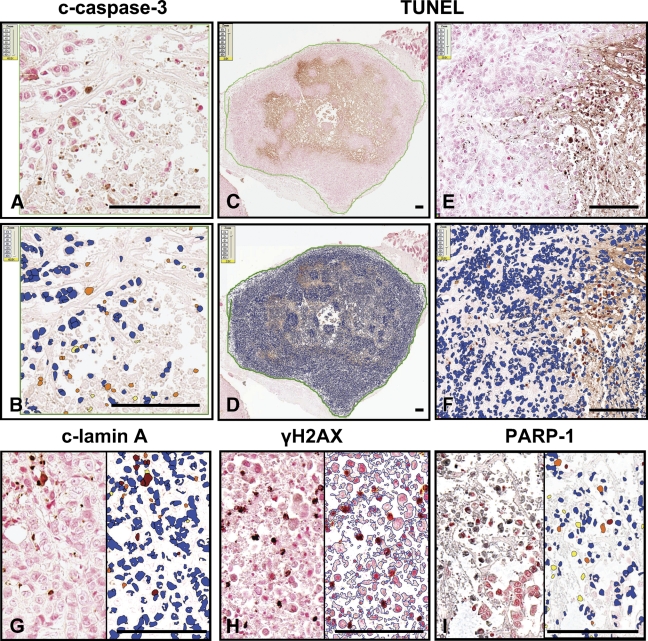
Figure 2.
Application of the nuclear algorithm to immunostained cancer xenograft specimens. The nuclear algorithm was applied to breast tumor specimens immunostained for cleaved caspase-3 (A,B), cleaved lamin A (G), γH2AX (H), PARP-1 (I), and labeled by TUNEL assay (C–F). On the digital slide images (A,C,E, and left side of G–I), brown and black colors denote immunopositive staining (brown, DAB chromogen; black, SG chromogen). Specimens were counterstained with Nuclear Red. In the mark-up images (B,D,F, and right side of G–I), red, orange, and yellow pixels visualize immunopositive nuclei (strong, moderate, and weak intensity, respectively), whereas blue pixels depict immunonegative nuclei. Bar = ∼100 μm.

