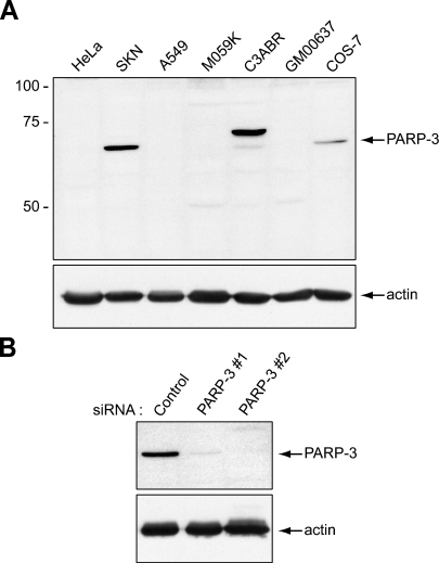
Figure 1.
Validation of the specificity of the poly(ADP-ribose) polymerase 3 (PARP-3) antibody by Western blot analysis. (A) Top panel: The anti–PARP-3 antibody detects a single protein band of 62 kDa in the human cell line SK-N-SH and in the monkey cell line COS-7. In the C3ABR human cell line, both PARP-3 isoforms, of 67 kDa and 62 kDa, are detected (see text). The other cell lines do not express detectable levels of PARP-3. Lower panel: A duplicate blot was probed with an anti-actin antibody to show equal loading. (B) Top panel: In SK-N-SH cells transfected with small interfering RNAs (siRNAs) targeting PARP-3 (PARP-3#1 or PARP-3#2), levels of the 62-kDa protein detected with the PARP-3 antibody are drastically reduced. Levels of the 62-kDa protein are not affected by transfection with a control siRNA. Lower panel: A duplicate blot was probed with an anti-actin antibody to show equal loading.

