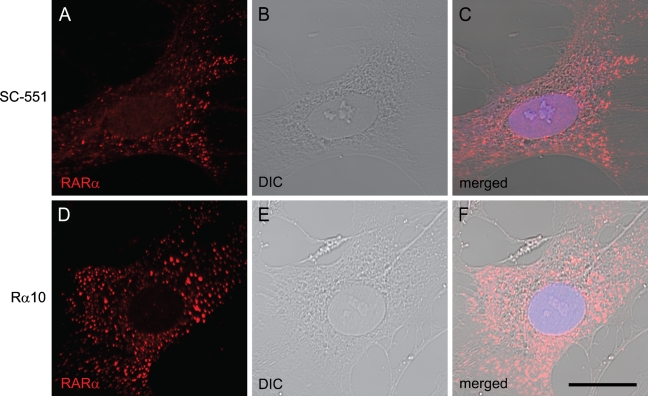
Figure 1.
Cytosolic speckled distribution of retinoic acid receptor alpha (RARα) proteins in activated hepatic stellate cells (HSCs). Primary HSCs collected from rat liver were spontaneously activated by culturing on poly-l-lysine–coated glass-bottom dishes. After 7 days of culture, cells were fixed and stained by rabbit polyclonal anti-RARα antibody (A–C) or mouse monoclonal anti-RARα antibody (D–F). RARα localization is shown in red (A,D). Differential interference contrast (DIC) images (B,E) and merged images (C,F) are also shown. Bar = 20 μm and applies to all the panels.

