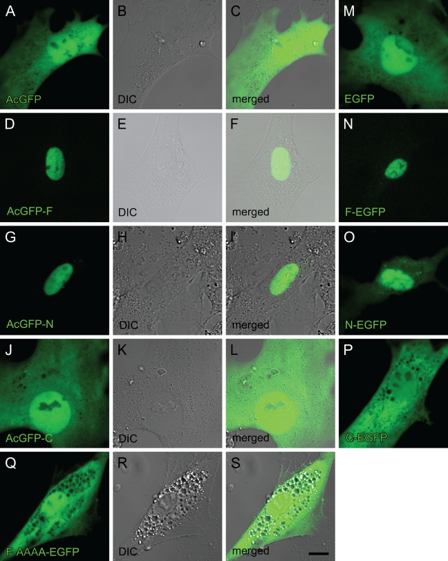
Figure 4.
Cellular localization of RARα protein fragments or a mutant fused with GFP in activated primary rat HSCs. Cells were transfected with GFP-expressing plasmids inserted with cDNA for RARα fragments or a mutant designated in Figure 2 3 days after seeding. The position of the GFP tag is either at the N terminus (A–L) or the C terminus (M–S). Four days after transfection (7 days after seeding), cells were observed by confocal laser scanning microscopy. GFP fluorescence is shown in green (A,D,G,J,M–Q). DIC images (B,E,H,K,R) and merged images (C,F,I,L,S) are also shown. Bar = 10 μm and applies to all the panels.

