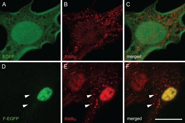
Figure 5.
Cellular localization of exogenous and endogenous RARα proteins in activated primary rat HSCs. Cells were transfected with GFP-expressing plasmids with (D–F) or without (A–C) cDNA for full-length RARα 3 days after seeding. Four days after transfection (7 days after seeding), cells were fixed and stained by rabbit polyclonal anti-RARα antibody. GFP fluorescence, which represents exogenous RARα protein localization, is shown in green (A,D). Staining by anti-RARα antibody, which recognizes both endogenous and exogenous RARα proteins, is shown in red (B,E). Merged images are also shown (C,F). Arrowheads indicate cytosolic dots stained in red but not in green, indicating that these cytosolic dots are composed solely of endogenous RARα proteins. Bar = 20 μm and applies to all the panels.

