
Figure 1.
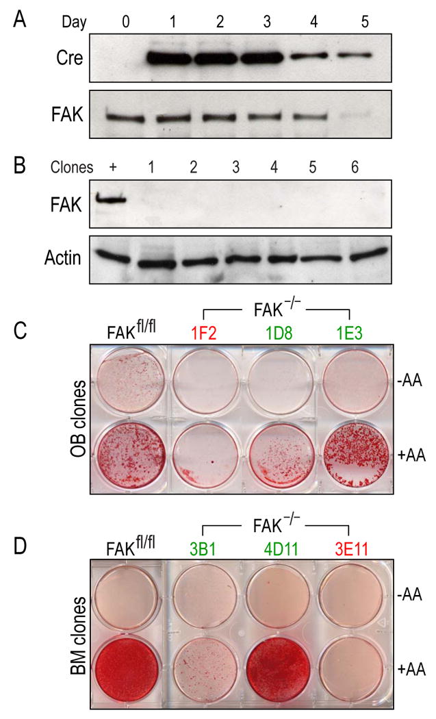
Establishment of FAK null osteoblast and bone marrow cell lines. (A) In vitro knockout of fak in osteoblasts. Cells from floxed animals (FAKfl/fl;p53−/−) were treated with adenovirus carrying cre. One microgram of total protein lysates was analyzed by Western blot using a monoclonal antibody against Cre. Signal was detected using enhanced chemiluminescence. After 5 days, the Cre protein level was tapered off. The same lysates were subjected to Western blot using a monoclonal antibody against FAK. FAK level was significantly reduced after 5 days of infection. (B) Establishment of FAK null osteoblast clonal lines. One microgram of total protein from immortalized calvarial osteoblasts was analyzed using monoclonal antibody against FAK. β-actin was used to ensure an equal amount of protein loading. +; positive control (FAKfl/fl;p53−/−), lane 1-6; FAK null osteoblast clonal lysates. (C,D) Calvarial osteoblast (C) and bone marrow cell line (D) differentiation in vitro. Immortalized calvarial FAK null clones were induced to differentiate in vitro. Cells were grown to confluency and differentiation was induced using ascorbic acid and β-glycerophosphate. After 2 weeks, cells were stained with 0.2% Alizarin Red to detect mineralization. A portion of FAK null clones (30%) differentiated in vitro. Mineralized and non-mineralized clones were labeled in red and green respectively. FAKfl/fl; parental cell line (FAKfl/fl;p53−/−), −AA; uninduced, +AA: induced by ascorbic acid and β -glycerophosphate; OB: osteoblast; BM; bone marrow.