
Figure 3.
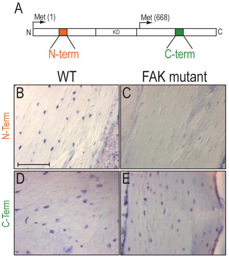
In situ hybridization for FAK in bone tissues. (A) Diagram of FAK protein and cDNA probe regions. FRNK is initiated at a methionine 668 and results in a truncated FAK protein. (B-E) In situ hybridization for N-terminal (B,C) and C-terminal of FAK transcripts (D,E) in wildtype and FAK mutant tibiae. FAK mutants were void of the transcripts for the N-terminal of FAK (C), while they expressed transcripts for the C-terminal of FAK transcripts (E). Bar: 100 μm.