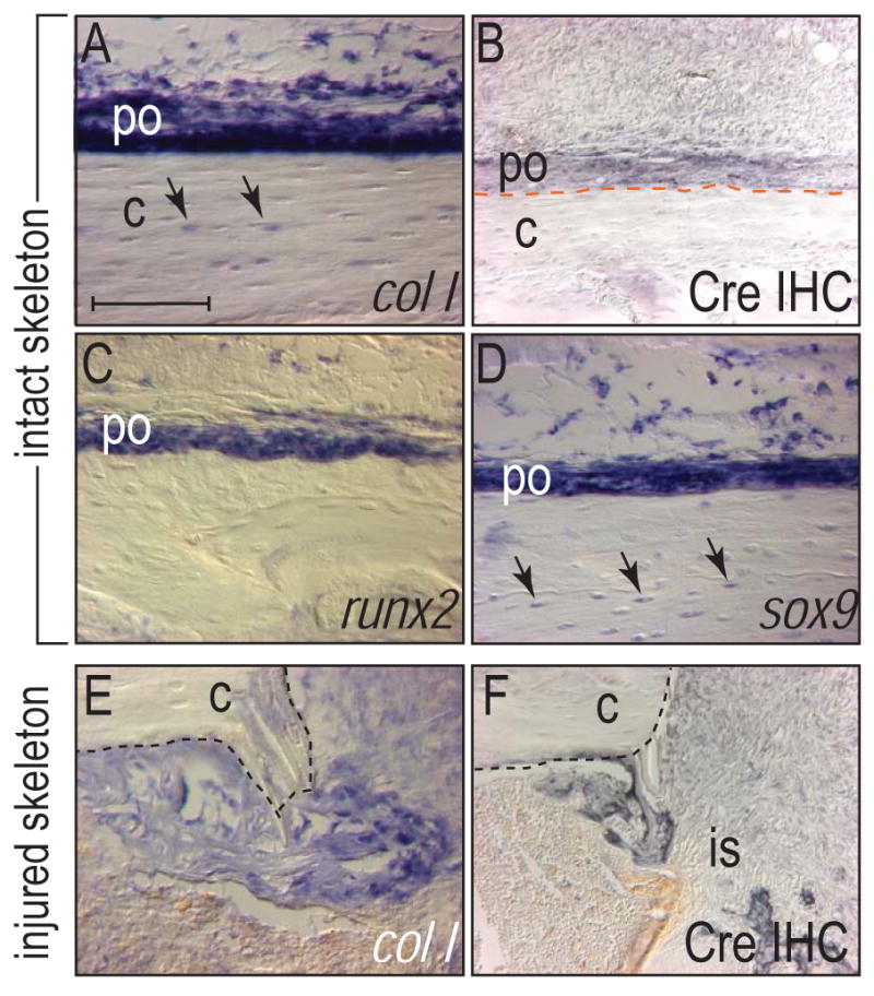
Figure 4.

Molecular analyses of osteogenic genes in wild type animals and Cre expression in the Col1-Cre mouse. (A) In situ hybridization of wild type tibia for collagen type I. Col I was expressed in osteocytes (arrows) and in the cambial layer of the periosteum. (B) Immunohistochemistry for Cre in the tibia of the Col1-Cre mouse. Cre protein was detected in the periosteum. (C,D) In situ hybridization for runx2 (C) and sox9 (D) in wild type tibia. Sox9 was expressed in both periosteum and osteocytes (arrows). (E,F) Collagen type I (col I) in situ hybridization and Cre immunohistochemistry in the pinhole injury sites from FAK mutants at d7. Col I and Cre expression was colocalized in the injury site. (E). c: cortex, is: injury site, po: periosteum. Bar: 100 μm.
