
Figure 5.
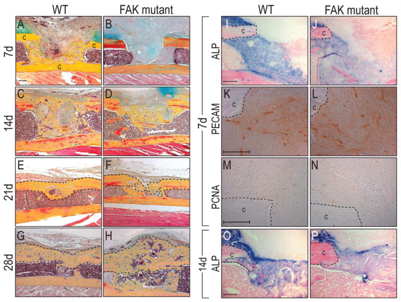
Delayed bone healing in FAK mutants. Mono-cortical tibial injuries were induced in wild type and FAK mutants. (A-H) Histology of wild type and FAK mutant injury at post-surgical d7, 14, 21 and 28. FAK mutants showed delayed skeletal healing compared to wild type animals. At 28 days after surgery, FAK mutants developed exuberant calluses compared to wild type counterparts. (I,J) Alkaline phosphatase activity in the injury sites at post-surgical d7. FAK mutants (J) showed less enzyme activity in the injury site compared to wild type animals (I). (K,L) PECAM staining at post-surgical d7 showed equivalent number of endothelial cells in both wild type (K) and FAK mutant (L). (M,N) PCNA staining revealed a similar proliferative activity in wild type and FAK mutant. c: cortex, is: injury site. Bar: 100 μm.