Figure 7.

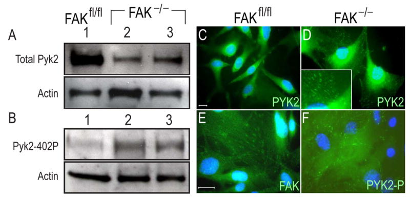
Western and Immunofluorescence analyses of FAK and Pyk2 in FAK null osteoblast cell lines. (A) Western blot of total Pyk2 protein in FAK null osteoblast clones (FAK−/−;p53−/−). A total of 2 μg of cell lysate was loaded in each lane. Membrane was blotted with Pyk2 antibody (A) and phosphospecific Pyk2 antibody (B). β-actin was used for a loading control. The activated form of Pyk2 was increased in FAK null cells. (C,E) Immunofluorescence of total Pyk2 (C) and FAK (E) in wild type (FAKfl/fl;p53−/−) osteoblast cells. Pyk2 was found in the perinuclear region in wild type cells. (D,F) Immunofluorescence analyses of total Pyk2 (D) and phosphospecific Pyk2 (F) in FAK null osteoblast cells (FAK−/−;p53−/−). Inset in panel D; high magnification of focal adhesions. When FAK was deleted, Pyk2 was redistributed into focal adhesions in FAK null cells. Active form of Pyk2 was found in focal adhesions in FAK null cells. Bar; 10 μm.
