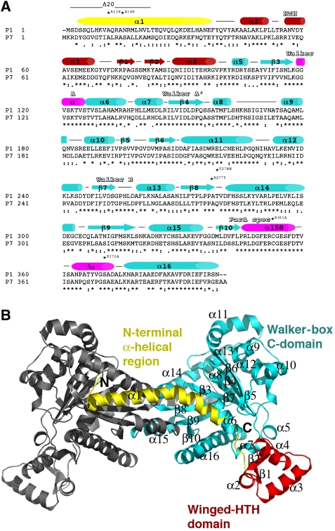
Figure 1.
Sequence homology of P1 and P7 ParA proteins and overall structure. (A) Sequence alignment of the P1 and P7 ParA proteins. Secondary structural elements are indicated over the sequences and the three structural regions are coloured yellow (N-terminal α-helical region), red (winged-HTH region) and cyan (Walker-box C-domain). Regions that fold into helices on ADP binding are indicated by pink cylinders. The HTH, Walker A, Walker A′, Walker B and ParA-specific regions are labelled. Residues mutated in the study are also labelled. (B) Ribbon diagram of the P7 apoParA dimer. The molecule is coloured coded as in (A). This figure and Figures 2, 3A–C, 4A–C, 5 and 6A were made with PyMOL (Delano, 2002).

