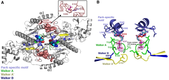
Figure 5.
ADP-binding interactions: Walker motifs and a second ADP-binding pocket. (A) Overall structure of the P212121 ParA–ADP structure. The α1 helices and the Walker motifs, A, A′ and B are labelled. The Walker motifs are coloured green (Walker A), yellow (Walker A′) and dark blue (Walker B). The ParA-specific motif is cyan. The second ADP-binding site is coloured salmon and the ADP is shown as CPK. Inset to the upper right is a close-up view of the interactions between the two ParA subunits and the second (non-primary)-bound ADP molecule. (B) Close-up of the ‘primary' ADP-binding site and a 2.05 Å resolution Fo–Fc map (magenta mesh), calculated after omitting the ADP, and contoured at 4.5σ. The nucleotide-binding motifs are coloured as in (A).

