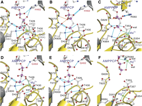
Figure 3.
Details of the triphosphate-binding site in the P-domain. MolA (A) and MolB (B)of CopA-PN wild type, those of His462Q mutant (D, E) and Ca2+-ATPase (C: E1·AMPPCP form (Toyoshima and Mizutani, 2004), F: E2·ATP(TG) form: PDB entry 2DQS). Side chains of important residues and AMPPCP are shown in ball-and-stick. Broken lines in light blue show likely hydrogen bonds, and those in green show coordination of the divalent cation. Broken lines in orange indicate hydrogen bonds specific to MolB (B) and Ca2+-ATPase (C). Small spheres represent Mg2+ (green), Me2+ (most likely Ca2+ in the crystal structure (Picard et al, 2007); cyan), and coordinating water molecules (red). Prepared with Molscript (Kraulis, 1991).

