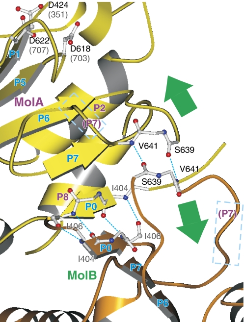
Figure 5.
Crystal contact between the P-domains of adjacent protomers. Dotted lines in cyan show hydrogen bonds between the β-strands in neighbouring protomers (MolA, yellow; MolB, orange). Rectangles in broken lines show the P7 helices, which has an important role in transmitting the binding signal of Mg2+ to the transmembrane domain in Ca2+-ATPase (Toyoshima and Mizutani, 2004). Green arrows indicate the movements expected for the part of the P-domain induced by the binding of Mg2+.

