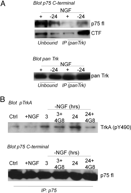
Fig. 4.
(A) Immunoprecipitation analysis performed with anti-pan-Trk (A) and p75 C-terminal antibodies (B) on lysates 24 h after NGF removal. Immunocomplex amounts were analyzed by immunoblotting with anti-p75 C-terminal (A). Note that to detect full-length p75 species (fl p75), longer time of exposure was needed than those necessary for CTF band. To demonstrate that equal amounts of protein were brought down after the immunoprecipitation procedure, membranes were stripped and reprobed with pan-Trk antibody. (B) Immunoprecipitation analysis performed with p75 C-terminal antibody (see Methods). Immunocomplexes were analyzed by immunoblotting with TrkA(pY490) antibody. The amount of p75 brought down by immunoprecipitation was analyzed by blotting with p75 C-terminal antibody (fl p75). Ctrl, samples before NGF exposure; +NGF, samples exposed to NGF; 3, 24, 24-h NGF-deprived samples; 3 + 4G8, 24 + 4G8, 24-h NGF-deprived samples incubated with Abeta antibody (MAb4G8).

