Fig. 2.
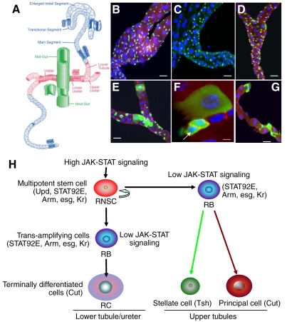
The Malpighian tubules (MTs) and renal stem cell lineage in Drosophila. (A) Drawing of the Drosophila MTs (modified from Wessing and Eichelberg, 1978). The adult MTs consist of two pairs of epithelial tubes: a longer, anterior pair runs through the hemolymph on both sides of the midgut, and a shorter, posterior pair runs along the hindgut. The pairs converge at a common ureter at the midgut–hindgut junction. Each tubule is divided into four compartments: initial, transitional, main and proximal (lower tubules and ureter). (B–D) Expression pattern of unique molecular markers in the region of lower tubules and ureters of adult Drosophila MTs. (B) Kr-Gal4/UAS-GFP is specifically expressed in the small nuclear cells (anti-GFP, green; anti-Arm, red; DAPI, blue). (C) upd-Gal4/UAS-GFP expressed in the region of the lower tubules and ureters (anti-GFP, green; DAPI, blue). (D) Stat92E-GFP reporter is expressed only in small nuclear cells in the region of the lower tubules and ureters (anti-Arm, red; anti-GFP, green; DAPI, blue). (E–G) MTs with GFP-marked wild-type MARCM clones. (E) Six days after clone induction, GFP marks clusters of cells with small, intermediate and large nuclei in the region of the lower tubules and ureters. (F) An enlarged view of a GFP-marked clone from E [arrow, renal and nephric stem cells (RNSCs), anti-arm, red; anti-GFP, green; DAPI, blue]. (G) 10 days after clone induction in the upper tubule, the GFP labels both stellate and principal cells. (H) Schematic summary of renal and nephric stem cell lineage in Drosophila MTs (modified from Singh and Hou, 2008). Markers expressed in RNSCs, and their differentiated cells are shown in parentheses. RB, renalblast; RC, renalcyte. Scale bars, 10 μm (B–E), 5 μm (F–G).

