. Author manuscript; available in PMC: 2009 Jun 21.
Published in final edited form as: Circulation. 2008 May 27;117(22):2919–2927. doi: 10.1161/CIRCULATIONAHA.107.754614

Figure 4.

Figure 4

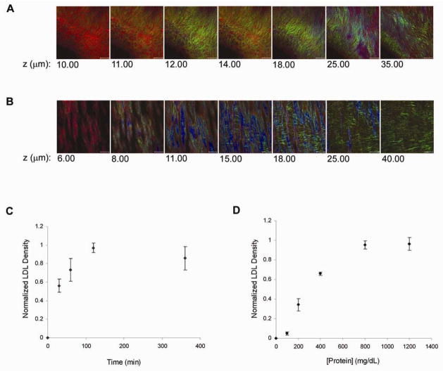
LDL binding along the porcine coronary arterial wall. A, Selected z-series two-photon images of a pig coronary arterial branch point. LDL (red) bound extensively along the surface of the exposed collagen fibrils (green). Scale bar, 50 μm. B, A minimal amount of LDL (red) bound to the surface of the IEL (blue). Scale bar, 50 μm. C, Saturation curve of LDL binding to branch points at various incubation times with 2 mg/mL LDL. LDL binding reached saturation after about two hours of incubation with the artery (n = 4). D, Steady-state LDL binding studies in PBS showed a sigmoid pattern where (n = 3):
Y=1/(1+e(x103)/301))