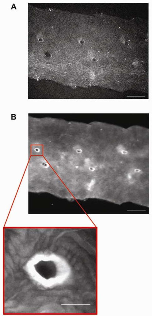
Figure 5.
LDL binding along the mouse aorta. A, Mouse aorta prior to incubation with LDL. Autofluorescence was low at 680 nm. Paired intervertebral branch points along the aortic wall became visible at a high intensity and increased exposure time (Intensity = 100%, exposure = 680 ms and gain = 165). Scale bar, 500 μm. B, Mouse aorta after incubation with LDL. LDL bound extensively to a circular region around intervertebral branch points. LDL bound lightly along the rest of the arterial wall (Intensity = 7.72%, exposure = 31 ms, Gain = 114). Scale bar, 500 μm. Zoom scale bar, 100 μm.

