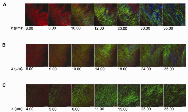
Figure 6.
LDL binding to pig coronary arterial branch points in various buffered solutions. A, LDL binding in dH2O was very high and LDL formed insoluble complexes along the surface of the exposed collagen. B, LDL binding in normal saline was lower than that seen in dH2O above or in bicarbonate buffer B. C, LDL in a highly anionic buffer composed of 1X PBS with 2 mM azide showed very low binding to the surface of the branch point. Scale bar, 50 μm.

