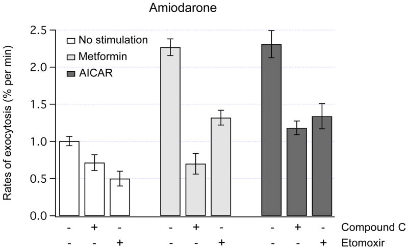
Fig. 3.
AMPK-dependent exocytosis in lipid-loaded cells. The rates of exocytosis were measured using FM1-43 fluorescence in amiodarone-treated HTC cells. Amiodarone increases intracellular lipid content in HTC cells by ~ 2-fold [11]. Acute exposure to metformin (1 mM) or AICAR (0.5 mM) potently increased the rates of exocytosis (P < 0.001 for both). Pretreatment with 20 μM Compound C (5 min) to inhibit AMPK did not significantly change the rates of exocytosis under basal conditions, but markedly decreased the rates of exocytosis evoked by metformin or AICAR (P < 0.001 for both). Pretreatment with R-etomoxir (100 μM, 1 hour) to inhibit FAO decreased the rates of basal exocytosis (P < 0.006). Note that metformin and AICAR stimulated exocytosis even in the presence of R-etomoxir (P < 0.01 for both). The number of cells analyzed was from 7 to 46.

