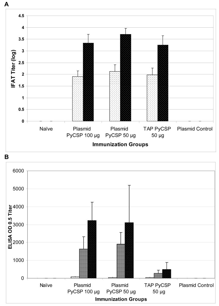
Figure 4. Antibody responses against sporozoite stage antigens.
BALB/c mice were immunized IM with 100 or 50 μg plasmid DNA or 50 μg of TAP fragments encoding PyCSP or empty VR1012 plasmid (plasmid control) 3 times at 3-week intervals. Sera were collected 2 wk after each immunization. Control sera were collected from unimmunized naïve mice. Sporozoite-specific antibodies were assayed by (A) Indirect Fluorescent Antibody Test (IFAT) against P. yoelii sporozoites using pooled sera (n=4/group) or (B) ELISA against recombinant PyCSP capture antigen, using sera collected post first (white bars with black dots), second (white bars with black stripes; shown for ELISA only) or third (black bars with white dots) immunizations. Histograms represent (A) logarithmically transformed geometric mean IFAT +/− standard deviation or (B) geometric mean ELISA OD 0.5 +/− standard deviation of pooled sera.

