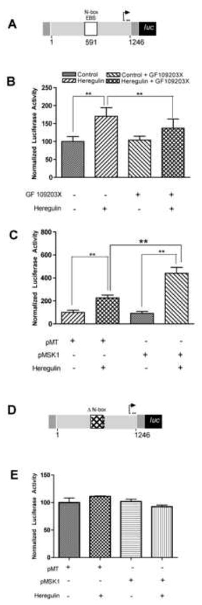
Figure 2. MSK1 plays a critical role in heregulin-induced utrophin-A promoter activation.

Schematic (A) of the utrophin-A promoter luciferase construct was cotransfected into C2C12 cells along with transfection control pRL-TK. C2C12 cells transfected with utrophin-A promoter-reporter were serum starved and (B) treated with 2nM heregulin and the MSK inhibitor (5μM of GF109203X) for 30′ as indicated and showed significant reduction of heregulin-induced utrophin-A promoter activity in the presence of pharmacological inhibition of MSK1/2. (Bargraph: control 100±13.73; HRG 170.30±24.22; control+GF109203X 104.2±10.65; HRG+GF109203X 137.3±25.47; n=5). These cells were also (C) transfected with either the empty vector pMT or MSK1 expressing constructs (pMSK1) prior to overnight serum starvation and heregulin stimulation and showed potentiation of heregulin-induced utrophin-A promoter activity by MSK1. (Bargraph: pMT 100.0±19.28; pMT+HRG 227.10±23.04; pMSK1 91.71±17.41; pMSK1+HRG 440.50±51.65; n=5). Schematic (D) of the N-box deleted utrophin-A promoter luciferase construct cotransfected into C2C12 cells along with pRL-TK. Cells were also (E) transfected with either empty vector pMT or MSK1 constructs (pMSK1) prior to overnight serum starvation and heregulin stimulation. Neither heregulin nor MSK1 showed upregulation (Bargraph: pMT 100.9±14.58; pMT+HRG 106.5±1.686; pMSK1 102.7±7.617; pMSK1+HRG 93.27±5.339; n=5). Luciferase activity is normalized to pRL-TK-derived luciferase activity (internal control) and expressed as 100% in the control group. ** statistical significance p<.01
