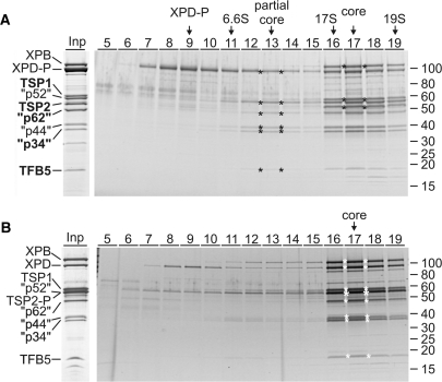
Figure 1.
Characterization of T. brucei TFIIH. (A) The input lane (Inp) shows the final eluate of a XPD–PTP purification separated by SDS–PAGE and stained with Coomassie. On the left, the TFIIH subunit orthologs are specified (quotation marks indicate that the sizes of the trypanosome proteins are different). The five new identifications are indicated by bold lettering. An equivalent eluate was sedimented through a linear 10–40% sucrose gradient which was fractionated from top to bottom. Proteins from each fraction were separated by SDS–PAGE and stained with Sypro ruby. The six proteins of the partial core and the three additional proteins of the TFIIH core complex are marked by asterisks in their peak fractions. Marker proteins with known S-values were analyzed in parallel gradients. Protein marker sizes are specified on the right. (B) A corresponding analysis is shown for TSP2-PTP purified material. Due to the tag, TSP2-P co-migrates with the p52 orthologue (large asterisk).

