Figure 5.
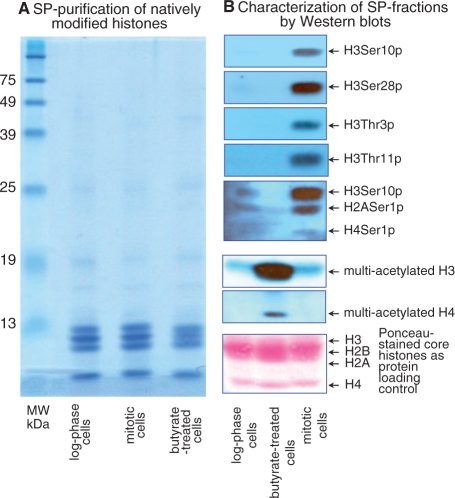
SP-purification of hyper-phosphorylated and hyper-acetylated core histones from HeLa cells extracted with H2SO4. (A) Coomassie-stained SDS–PAGE of SP-eluted core histones from logarithmically growing HeLa cells (lane log phase cells), metaphase-arrested cells enriched in hyper-phosphorylated core histones (lane mitotic cells) and butyrate-treated cells enriched in hyper-acetylated core histones (lane butyrate-treated cells). (B) Immunoblotting analysis of SP-eluted core histones using antibodies to the most prominent hyper-phosphorylated residues during mitosis (lane mitotic cells), and to globally hyper-acetylated histones H3 and H4 from butyrate-treated HeLa cells (lane butyrate-treated cells) (note that butyrate-treated lane and mitotic cell lane are inverted with respect to the same lanes in the Coomassie-stained gel). The lane log phase cell in (B) is a blotting-control of SP-eluted core histones from logarithmically proliferating HeLa cells; these histones are largely hypo-phosphorylated and hypo-acetylated. The bottom lower panel (in red) is the same membrane stained with Ponceau S, showing transferred core histones, prior to immunoblotting, and serves as a protein loading control.

