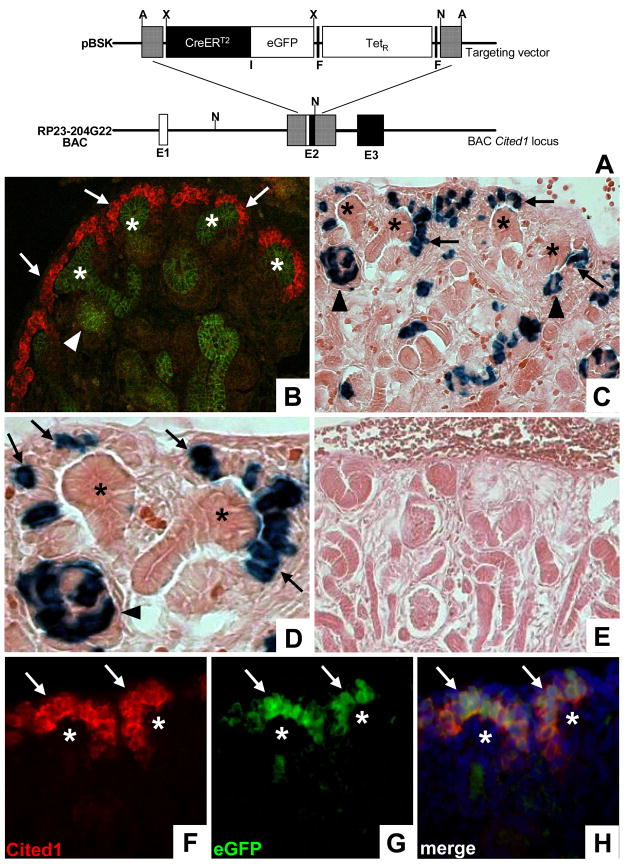
Figure 1. Generation and characterization of Cited1-CreERT2 mice.
A, Schematic of the targeting strategy used to create the BAC Cited1-CreERT2-IRESeGFP transgene. Hatched lines represent regions of homology. Following BAC targeting TetR was removed through Flp mediated recombination E1–Exon 1, F–Frt sites, I–IRES; A–Afl II, N–Not I, X–Xho I. B, Dual immunofluorescence staining of E15 mouse kidneys using anti-Cited1 (red) and anti-ECadherin (green) antibodies demonstrates endogenous Cited1 expression in the cap mesenchyme (arrows), and the absence of Cited1 expression in all ECadherin positive epithelial structures including renal vesicles (arrowhead) and UBs (asterisk). C/D, Low (C) and high (D) power images of the nephrogenic zone of E18 β-Gal stained Cited1-CreERT2/R26RLacZ kidneys following E15 maternal injection of tamoxifen. Recombination is observed within the cap mesenchyme (arrows) and early nephronic epithelia including renal vesicles (arrowheads). No recombination is observed in UBs (asterisks). E, β-Gal stained kidney from E18 Cited1-CreERT2/R26RLacZ embryo whose mother was not injected with tamoxifen. F/G, Anti-Cited1 antibody staining (F) corresponds to eGFP signal (G) within the cap mesenchyme (arrows) in mice carrying the Cited1-CreERT2-IRESeGFP transgene. No expression observed in UBs (asterisk). H, Merged image of (F) and (G) demonstrating overlapping expression of Cited1 and eGFP.

