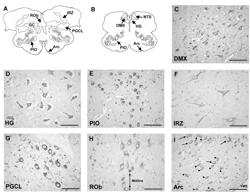
Figure 3.
5-HT2A receptor immunostaining in the human infant medulla at 10 postnatal months. Panels (A) and (B) show diagrams of horizontal sections of rostral and mid-medulla, respectively, showing the location of the component nuclei of the medullary 5-HT system at each level. 5-HT2A receptor immunostaining is punctate and localized to neuronal cell bodies and processes, indicative of post-synaptic localization of the receptors. Figure shows intense immunostaining of neurons in the DMX (C), motor neurons in the HG (D), and spherical neurons in the PIO (E). Moderately stained neurons of heterogenous morphology were observed in the extra-raphé regions including the GC (F). Spherical 5-HT2A immunoreactive neurons were observed in the lateral extent of the PGCL (G) consistent in location and morphology with preBötC neurons in rodents. Lightly stained spherical and fusiform 5-HT2A receptor immunoreactive neurons were observed in the midline raphé (H). Midline of the medulla is labeled by double headed arrow; D, dorsal; V, ventral. Immunostaining to “classical” arcuate neurons (arrows) and astrocytes (arrow-heads) in the arcuate nucleus (I). VMS, ventral medullary surface. All images at ×40. Scale bar= 100 μm.

