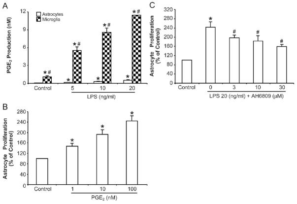
Figure 3. PGE2 is involved in astrocyte proliferation.
(A) Primary enriched astrocyte and microglial cultures were treated with LPS at given doses. The supernatant were collected 72 h after treatment and PGE2 concentrations were assayed using an ELA kit as described in Material and Methods. * p <0.05, compared with corresponding control cultures; # p <0.05, compared with astrocytes after same treatment. (B) Different concentrations of PGE2 were added to purified astrocyte cultures and the incubated for 72 h. Cell proliferation was assayed using a BrdU ELISA kit. * p <0.05, compared to corresponding control cultures. (C) Astrocytes and microglia co-cultures in transwell were pre-treated with the non-selective PGE2 receptor antagonist AH6809 for 1 h then challenged with 20 ng/ml LPS for 72 h. Astrocyte proliferation was then measured. Results are mean ± S.E.M of three experiments performed in triplicate. * p <0.05, compared with control cultures; # p <0.05, compared with LPS treatment group.

