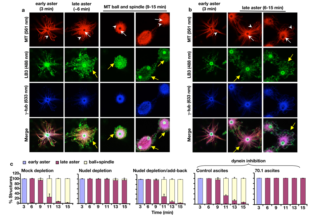
Figure 4.
Effects of disrupting Nudel or dynein on spindle assembly and LB3 localization in the AurA-bead based assay. (a) Progression of spindle morphogenesis and LB3 assembly in mock-depleted egg extracts. LB3 was found along MTs of early asters. As early asters transformed into late aster (white arrows and arrowheads point to bright MT cores and astral MTs, respectively), meshworks of LB3 were seen to surround the late asters. High levels of LB3 were also found throughout the MT balls and spindles. The bright green staining of the beads was caused by the rabbit secondary antibody that recognized anti-AurA antibodies coated on the beads. Yellow arrows point to the LB3 network surrounding the late aster, MT ball, and spindle. (b) Progression of MT and LB3 assembly in Nudel-depleted egg extracts. Formation of early and later asters and the accumulation of γ-tubulin on the AurA-beads occurred normally. However, there was a lack of MT ball and spindle assembly in the absence of Nudel. White arrows and arrowheads point to MT cores and astral MTs, respectively. There was a diminished LB3 network surrounding the late asters as compared to the late aster in the control (compare yellow arrows in the late asters in a and b). (c) Quantification of MT structures at different time points and treatment conditions. All images were acquired on a Leica SP5 confocal microscope with laser lines used indicated in parentheses. Scale, magnetic beads (2.8 µm in diameter). Error bars, standard deviation from >3 independent experiments.

