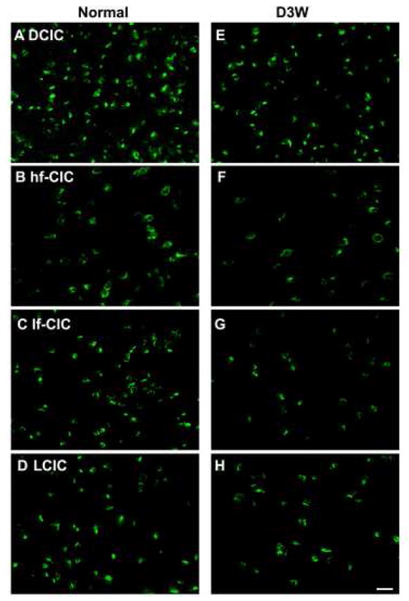
Figure 5.

Photomicrographs of representative in situ hybridization labeling of cells for TASK-1 in four regions of the rat inferior colliculus in normal hearing animals (Normal) (A-D)and in animals assessed three weeks following deafening (D3W) (E-H)from bilateral cochlear ablation in the Dorsal Cortex (DCIC) (A,E); high frequency region of the Central Nucleus of the Inferior Colliculus (hf-CIC) (B,F); low frequency region of the Central Nucleus of the Inferior Colliculus (lf-CIC) (C,G); lateral (external) cortex of inferior colliculus (LCIC) (D,H), scale bar = 25 μm.
