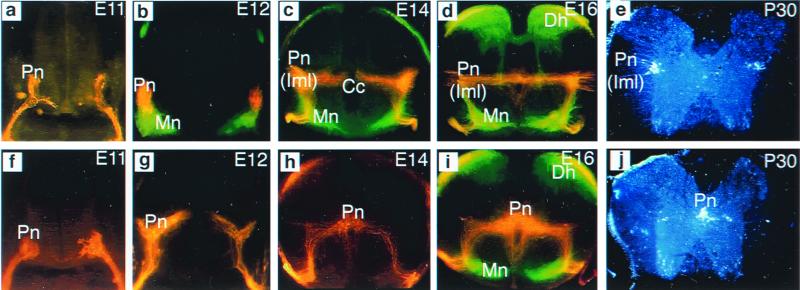
Figure 2.
Migratory pathways of sympathetic preganglionic neurons in upper thoracic spinal cord of wild-type and reeler mutant. Preganglionic neurons in embryos were retrogradely labeled with DiI crystals applied to the sympathetic ganglia; somatic motor neurons were retrogradely labeled with DiO crystals applied to the spinal nerves (a–d and f–i). Preganglionic neurons in postnatal mice were labeled by i.p. injection of Fluorogold (e and j). (a) E11 wild type. Transverse section shows that preganglionic neurons (Pn) have undergone their primary migration from the neuroepithelium to the ventrolateral spinal cord. (b) E12 wild type. Many preganglionic neurons (red) have migrated dorsally to separate from the somatic motor neurons (Mn, green). (c and d) E14 (c) and E16 (d) wild type. The majority of preganglionic neurons have completed their dorsal migration to arrive at their final location in the IML (Iml); a small number of neurons become localized to areas between the IML and the central canal (Cc). Note that in these micrographs, DiI labeling is found spanning the middle of the spinal cord as well as around the central canal. However, microscopic examination at higher magnification revealed that this is mostly fiber labeling. (e) Fluorogold labeling in a P30 mouse shows that the majority of sympathetic neurons are indeed located in the IML. (f) E11 reeler. Preganglionic neurons migrated, as in the wild-type mouse, to the ventrolateral spinal cord. (g) E12 reeler. Many preganglionic neurons stream toward the central canal, instead of migrating toward the IML as they normally do. The secondary migration of preganglionic neurons in the reeler mutant is, therefore, abnormal. (h and i) E14 (h) and E16 (i) reeler. The majority of preganglionic neurons have arrived at their final location adjacent to the central canal; a few neurons remain in the IML. Dh, dorsal horn. (j) Fluorogold labeling in a P30 reeler mutant confirms that the majority of preganglionic cell bodies are located adjacent to the central canal.

