
Figure 6.
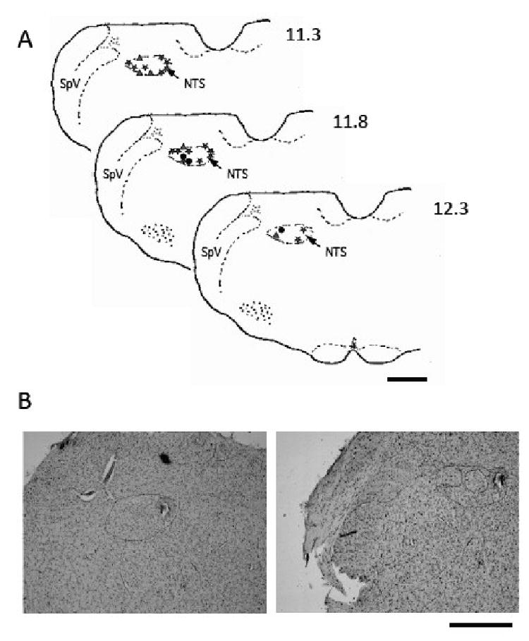
Location of cells recorded from the rostral NTS. A. Drawing of coronal sections of the brainstem showing the sites of electrolytic lesions indicating the location of NTS cells from which data were collected. Symbols are: star, taste-responsive cells; triangle, taste-responsive and non-taste-responsive cells recorded from approximately the same location; circle, non-taste-responsive cells. Numbers on upper right of drawing of each section indicate distance caudal to bregma. Abbreviations are: NTS, nucleus of the solitary tract; SpV, spinal nucleus of the trigeminal nerve. B. Photomicrographs of rostral NTS lesions showing the location of one NTS cell. Dotted line surrounds the NTS. Bars on lower right of each panel indicate 1.0 mm.