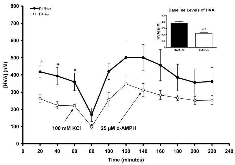
Fig. 5.
Graph showing extracellular measures of HVA resulting from reverse microdialysis with KCl or d-AMPH stimulation in the Str/NAc of D4R−/− and D4R+/+ mice. Although HVA significantly decreased in both genotypes as a result of KCl treatment, there were no significant differences in the amount of HVA reduction between genotypes. At the individual time points of 20, 40, and 60 minutes, baseline levels of HVA were significantly lower in D4R−/− mice (#p<0.05); the inset shows that combined baseline HVA levels were significantly lower in the D4R−/− mice (***p< 0.001). The error bars represent S.E.M.

