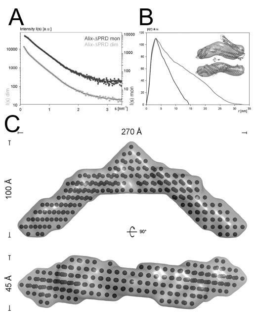
Figure 2. Small angle X-ray scattering analysis of ALIXBro1-V.
(A) Experimental scattering intensity patterns obtained for monomeric (dark grey) and dimeric (light grey) ALIXBro1-V are shown as a function of resolution and after averaging and subtraction of solvent scattering. The scattering intensity patterns calculated from the ALIXBro1-V monomer and dimer SAXS model with the lowest χ value are shown as black lines.
(B) P(r) function of both monomeric (dark grey) and dimeric (light grey) ALIXBro1-V (both curves have been adjusted to the same height). The ab initio model envelope of monomeric ALIXBro1-V is shown together with the manually docked ALIXBro1-V structure (Fisher et al., 2007) as inset.
(C) Ab initio modeling of dimeric ALIXBro1-V reveals a crescent shape model spanning ~ 270 Å; two orientations of the bead model including the molecular envelope are shown.

