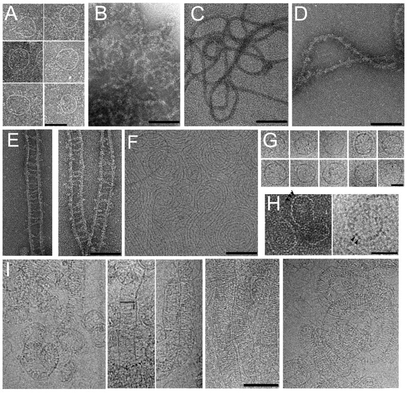
Figure 9. Electron microscopy analyses of CHMP4BΔC-ALIX and ALIXBro1-V complexes.
Negative staining EM images of (A) MBP-CHMP4BΔC, (B) MBP-CHMP4BΔC-ALIX, (C) CHMP4BΔC-ALIX, (D) CHMP4BΔC-ALIX - ALIXBro1-V (monomer) complexes, (E) CHMP4BΔC-ALIX- ALIXBro1-V (dimer) complexes. (F) Cryo-electron microscopy image of CHMP4BΔC-ALIX. (G) Cryo EM images of selected ring structures of CHMP4BΔC-ALIX; (H) Ring-like structures revealing the potential repeating unit, marked by arrows (left panel, negative staining, right panel cryo image). (I) 5 panels of cryo EM images showing CHMP4BΔC-ALIX - ALIXBro1-V (dimer) complexes. The length of one rung and the distance between rungs are indicated schematically in panel 2. The scale bars are 50 nm (A), 100 nm (B-F, I) and 30 nm (G, H).

