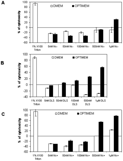
Figure 1. Cytotoxicity of increasing concentrations of ON705 delivered with Nx−40 (A), DLS (B), or with Nx+20 (C).
HeLa pLuc/705 cells were incubated with the different formulations in serum-containing medium (white bars) or in optiMEM (black bars) for 24 h. ON concentrations complexed with the liposomes are indicated. The level of cytotoxicity was determined with the tetrazolium-based colorimetric cell proliferation assay and expressed as percent of the cytotoxicity level in treated cells in comparison to non-treated cells. Cells incubated with 1% X-100 Triton are used as positive control. Error bars show standard deviation (n = 3).

