
Fig.2. DFMO inhibits apoptosis by activating ERK1/2, integrin β3, Src and AKT.
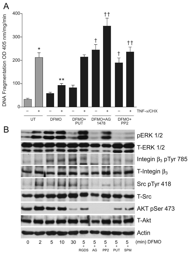
(A) Confluent serum starved cells were left untreated or pretreated with AG1478 (10 μM) or PP2 (10 μM) for 30 mins followed by DFMO (5 mM) or DFMO +putrescine (10μM) for 1h. These groups were then exposed to TNF-α/CHX for 3h. Cells were washed, and DNA fragmentation was measured as described in the methods section. (mean ± SE, n=3, p<0.05considered significant). *, significantly different from minus TNF-α/CHX UT, **, significantly different from TNF-α/CHX treated UT, †, significantly different from TNF-α/CHX treated DFMO group, ††, significantly different from TNF-α/CHX treated DFMO group or TNF-α/CHX treated UT group.
(B) Confluent serum starved cells were treated with 5 mM DFMO for the indicated time period. A second group of cells pretreated with RGDS or AG1478 or PP2 were treated with DFMO for 5 min. A third group of cells were treated with DFMO (5mM) +putrescine (10μM) or DFMO (5mM) + spermine (10μM) for 5 min. Cells were washed and lysed using lysis buffer containing protease and phosphatase inhibitors. Whole cell lysates were subjected to SDS-PAGE and western blot analysis using phospho-specific ERK1/2, Src, AKT, and integrin β3 antibodies. The membranes were stripped and probed with respective antibodies recognizing total protein. The membranes were also stripped and probed with β-actin antibody. Representative blots from 3 observations are shown.