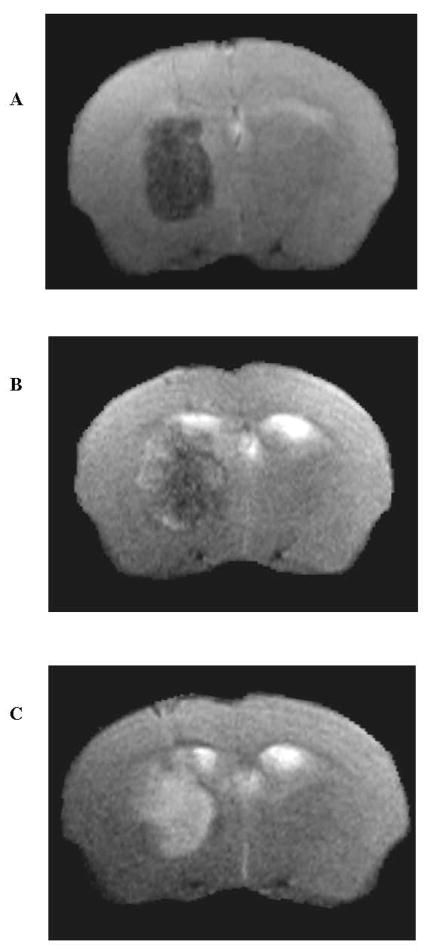
Figure 2.
A–C. MRI of APOE4TR mouse after collagenase-induced ICH at 2 h (A), 24 h (B), and 72 h (C) after injury. T2-weighted RARE spin echo images at 2 h (A) show predominantly low signal hematoma within the right basal ganglia, consistent with deoxyhemoglobin and intracellular methemoglobin. At 72 h (C), there is conversion to predominantly high signal, consistent with extracellular methemoglobin.

