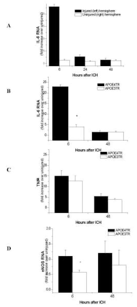
Figure 3.
A–CA–D. Quantitative q-PCR of inflammatory markers after ICH in APOETR mice. At 6, 24, and 48 h, IL-6 peaks and returns to normal levels in the injured hemisphere on APOE4TR mice when compared to the uninjured hemisphere (A). IL-6 (B) and eNOS (D) are significantly reduced in the injured hemispheres of APOE3TR mice compared to their APOE4TR counterparts. TNF-α was not significant(C).

