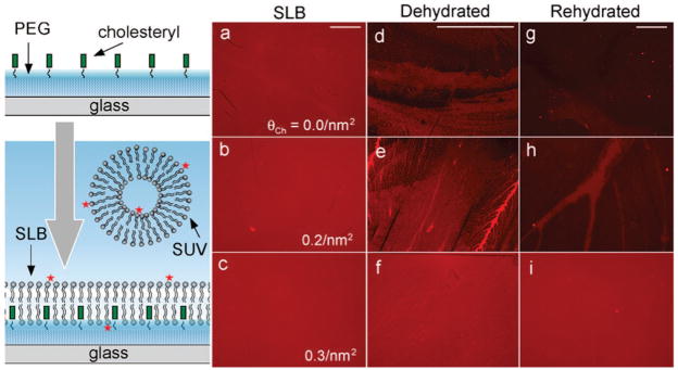
Figure 1.
Fluorescence microscopy images of supported lipid bilayers formed on a PEG brush surface with the indicated densities of surface tethered cholesteryl groups: θCh = 0.0 (a, d, g), 0.2 (b, e, h), and 0.3/nm2 (c, f, i). The scale bar (0.4 mm) for each column is shown at the top. The SLBs are formed from the fusion of SUVs containing 0.5% Texas-Red 1,2-dihexadecanoyl-sn-glycero-3-phosphoethanolamine (TR-DHPE), 50% egg phophatidylcholine (EggPC), and 50% 1,2-dioleoyl-3-trimethyl ammonium propane (DOTAP). Images for the as-formed SLBs in buffer solution (a, b, c) and those after dehydration (g, h, i) are taken with a 4× objective, while those in the dehydrated state (d, e, f) are taken with 10× objective to reveal more details.

