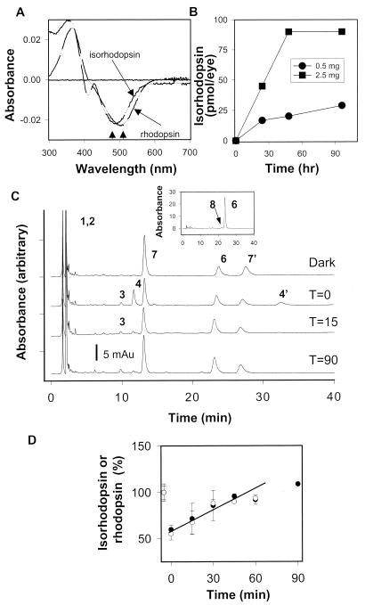
Figure 2.
Formation of isorhodopsin and kinetics of the retinoid flow in Rpe65−/− mice 48 h after 9-cis-retinal gavage. (A) Comparison spectra of rhodopsin from Rpe65+/+ mice and isorhodopsin from Rpe65−/− mice 48 h after 9-cis-retinal gavage (2.5 mg). Arrows denote differences in the absorption maximum for rhodopsin and isorhodopsin. (B) Isorhodopsin formation in Rpe65−/− mice at different time points after 0.5 mg or 2.5 mg of 9-cis-retinal gavage (n = 2). (C) Chromatograms illustrate the dark recovery of retinoids in Rpe65−/− mice 48 h after 9-cis-retinal gavage (2.5 mg) after a flash that bleached ≈45% of isorhodopsin. (C, Inset) isomeric composition of retinyl esters (8, 9-cis-retinol). (D) Dark recovery of isorhodopsin in Rpe65−/− mice 48 h after 9-cis-retinal gavage after a flash. For comparison, dark recovery of rhodopsin is shown for Rpe65+/+ (open hexagons) (7, 7′). anti- and syn- of 9-cis-Retinal oximes; all other peaks are as in the Fig. 1 legend.

