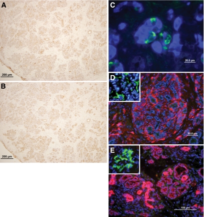
FIG. 4.
Histological examination of E42 pig pancreatic tissue 2–10 months after transplantation under the renal capsule of C57BL mice treated with the costimulatory blockade protocol (anti-LFA1, anti-CD48, and FTY720). Slides were stained for CD3+ lymphocytes (A), macrophages (F4/80) (B), insulin (blue) and glucagon (green) (C), mouse blood vessels (CD31, red), and pig blood vessels (CD31, green) (D), and cytokeratin (broad spectrum, red) and IgM and IgG deposits (green) (E). The inset in D demonstrates positive staining for pig endothelial cells (green) in E42 pancreas. The inset in E demonstrates positive normal staining for IgM and IgG deposits in the glomeruli of the kidney. The data are representative of the experiment shown in Table 1 (n = 11). (A high-quality representation of this figure is available in the online issue.)

