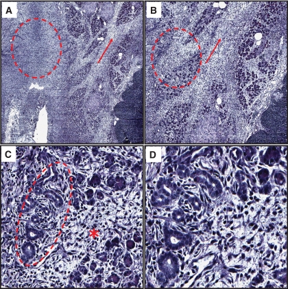
FIG. 6.
Necrotizing pancreatitis in a HIP rat treated with sitagliptin for 12 weeks. A: Representative image at ×2 magnification of the exocrine pancreas stained for hematoxylin and eosin from an HIP rat treated with sitagliptin for 12 weeks with necrotizing pancreatitis. Note partially preserved lobular configuration of the exocrine pancreas; however, note the significant loss of acinar cell density and the widening of the septae (arrow) as well as a complete loss of acinar cells in some areas (circle). B: Representative image at ×4 magnification. At this higher magnification, septal fibrosis and inflammation (arrows) are better appreciated as well as partial and complete loss of acinar cells (circle). C: Representative image at ×20 magnification. At this magnification, acinar cell injury and angulated tubular ductal structures within the acini are clearly seen (circle). Note the extensive septal inflammation and fibrosis (*). D: Representative image at ×40 magnification. At this higher magnification, angulated tubular ductal structures and surrounding tissue fibrosis are better appreciated. (A high-quality digital representation of this figure is available in the online issue.)

