Figure 3.
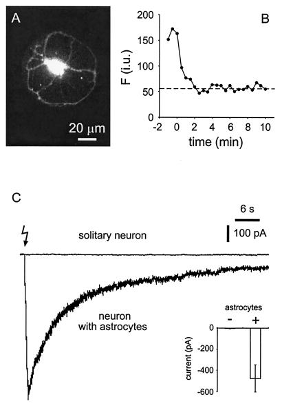
UV photolysis of NP-EGTA evoked an SIC in neurons cultured on astrocytic microislands but not in solitary neurons. Cells were coloaded with fluo-3 and NP-EGTA by bath incubation in the AM esters. To determine the period of dialysis necessary to remove fluo-3 and NP-EGTA from neurons, the fluorescent intensity of fluo-3 was monitored after establishing a whole-cell recording from a solitary neuron (A). In B, the intensity of fluorescence is plotted against time after establishing a whole-cell recording (t = 0). Note that fluorescence signal subsides until it reaches a level corresponding to autofluorescence (dashed line) well within the 10-min period that we adopted as a standard to dialyze NP-EGTA and fluo-3 from neurons in this study. i.u., intensity unit. (C) Neuronal currents recorded after UV illumination in the presence and absence of astrocytes. UV illumination, to photolyse NP-EGTA, caused an SIC in neurons that were cocultured with astrocytes (neuron with astrocytes) but not in solitary neurons. The lightning bolt indicates the onset of the UV stimuli (train of six UV pulses at 20 Hz). Inset summarizes all experiments (n = 8 for neurons with astrocytes; n = 5 for solitary neurons). Bars indicate means ± SEM.

