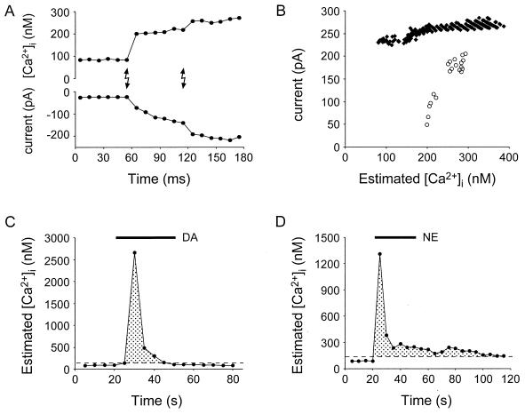
Figure 5.
Simultaneous recordings of astrocytic calcium levels and neuronal currents can be used to elucidate the relationship between astrocytic calcium levels and consequent glutamate release. UV pulses (A, lighting bolts) caused a step-like increase in astrocytic internal calcium (A, upper trace) and an increase in the inward current of adjacent neurons (A, lower trace). (B) The relationships between astrocytic calcium and SIC (and underlying glutamate release from astrocytes) for “all-or-none” (black diamonds) and “graded” (open circles) astrocytes are depicted. These photolysis experiments demonstrate that low levels of calcium are sufficient to induce a neuronal SIC. (C and D) The calcium responses of astrocytes to the application of dopamine (DA) and norepinephrine (NE) (both at 50 μM) demonstrate that the threshold calcium levels necessary to evoke neuronal SICs (dashed lines) are well within the range of the calcium levels that occur in astrocytes. Shaded regions represent astrocytic calcium levels that we have demonstrated are capable of stimulating glutamate release from these nonneuronal cells.

