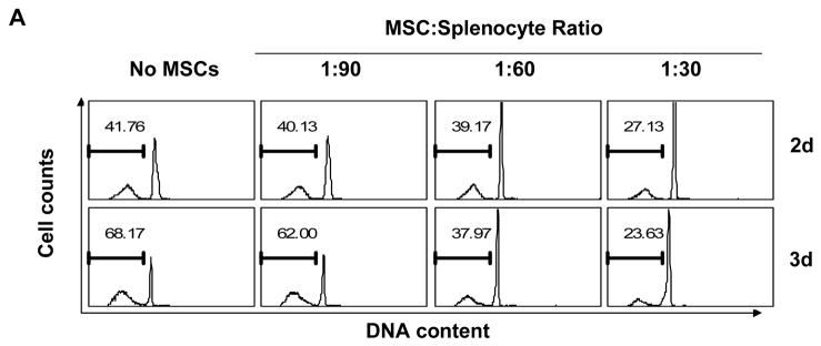
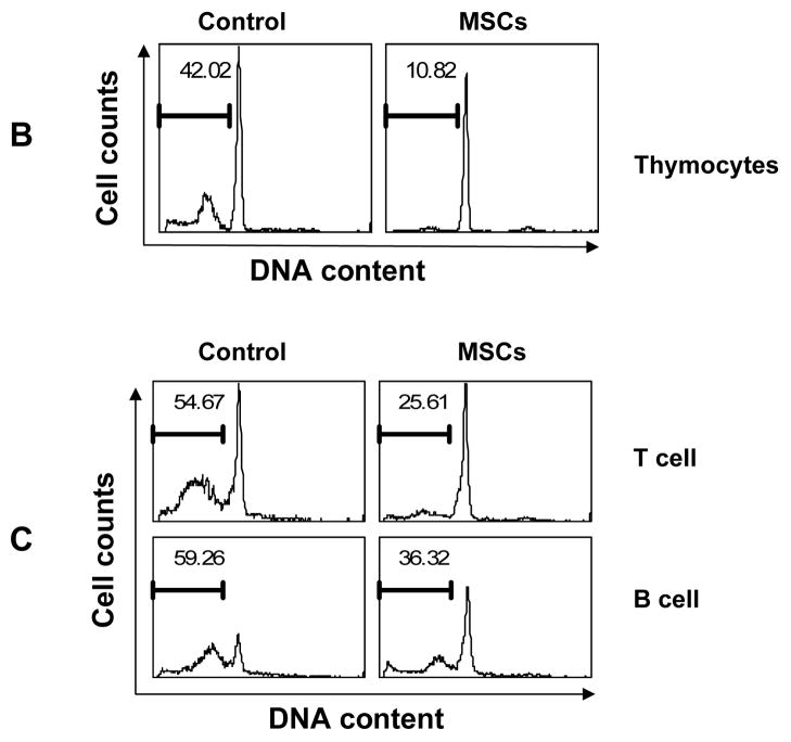
Figure 1. MSCs inhibit spontaneous lymphocyte apoptosis.
(A) Splenocytes were co-cultured with MSCs at a ratio of 30:1, 60:1 or 90:1 (splenocytes:MSCs), harvested at the indicated time points, then permeabilized and stained with propidium iodide to reveal hypodiploid peaks characteristic of apoptotic cells. (B) Thymocytes were co-cultured with MSCs at a 30:1 ratio for 2 days, and similarly analyzed for DNA content. (C) Purified T cells and B cells, prepared from splenocytes by immunoaffinity magnetic separation, were co-cultured with MSCs (30:1) for 2.5 days and DNA content determined. Values indicate percentage of cells with hypodiploid DNA content as detected by flow cytometry. Data shown are representative of four experiments.


