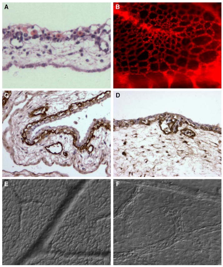
Fig. 1.
Ectoderm capillary plexus of day 12 chick embryo. a H&E staining. b Immunofluorescent staining with Lens culinaris agglutinin (LCA). c, d Immunohistochemical staining with endothelium-specific lectin Sambuco negro agglutinin (SNA). d DIC microscopy of live, non-fixed whole mounts of the CAM at the ectoderm plexus level (e) and mesoderm level (f), ×100 (a, c) and ×200 (b, d–f)

